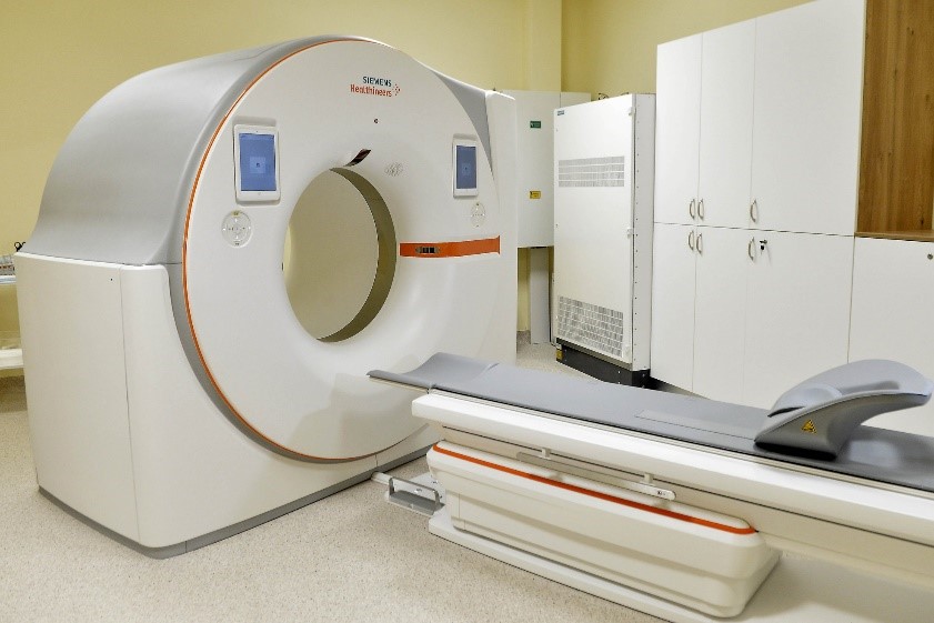
A CT device with a photon-counting detector, also new in the world, was handed over at Semmelweis University. Thanks to the equipment put into operation at the Medical Imaging Clinic, a faster and more accurate diagnosis can be expected, and the radiation dose - thanks to the photon counting technology - will be reduced by almost half.
At the ceremonial handover, Minister of Innovation and Technology László Palkovics said that this is the fourth time in the last seven years that one of the most modern imaging equipment in the world has been made available at the university. He reminded that Semmelweis University improved more than 140 places in the international ranking in one year.
Béla Merkely, rector of Semmelweis University, said that the university had reached a "real milestone", since equipment with such modern technology had not yet been put into operation in Europe. At one time, twenty such CTs were handed out around the world. He emphasized that the equipment works with a low radiation dose and is very accurate, which, in addition to benefiting patients, provides a new path in research.
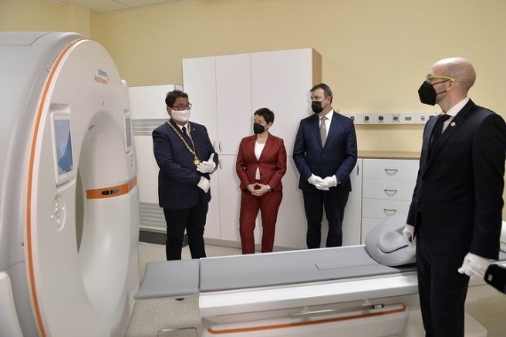
Pál Maurovich-Horvat, director of the Medical Imaging Clinic, presented the equipment and said that the Siemens Healthineers Naeotom Alpha CT device, thanks to its unique photon-counting technology, enables much more detailed spatial resolution compared to traditional CT, so even pathological changes of a few millimeters - the from tumors to calcified vessels and strictures - can also be identified with its help. He emphasized that the equipment is the fastest in the world and has the highest spatial resolution. This means that they can reach a more accurate diagnosis much faster than before, with lower radiation exposure.
In addition to grayscale images, the equipment is also suitable for taking color CT images based on tissue properties, thus enabling even more accurate diagnosis in oncology and cardiology, as well as lung, skull and joint imaging.
In oncological diagnostics, it is of great importance that the new CT, with a spatial resolution of 0.2 millimeters, is suitable for numerically determining the composition of lesions, so that benign and malignant tumors can be separated from each other with greater certainty.
Source: MTI
Photos: MTI/Lajos Soós

